PHẦN I: TÓM TẮT
PHẦN II: TẠM DỊCH
PHẦN I: TÓM TẮT KIẾN THỨC VỀ GAN NHIỄN MỠ LAN TOẢ
CÁC ĐẶC ĐIỂM CHUNG (General features):
- Gan to nhẹ (mild hepatomegaly) gặp trong 75% trường hợp
- Mức độ hút âm/tín hiệu(attenuation/signal) của gan chuyển sang của mỡ
- Vùng gan bảo tồn không bị nhiễm mỡ (focal fatty sparing):
- Xuất hiện dưới dạng các vùng dạng bản đồ (geographic regions)
- Vị trí đặc trưng (characteristic locations): Dọc theo hố/ giường túi mật (gallbladder fossa) HOẶC kế cận cửa gan/rốn gan (the porta hepatis) (hạ phân thùy IV: segment IV), dây chằng liềm (the falciform ligament) HOẶC nhu mô dưới bao gan (subcapsular parenchyma)
- Không có hiệu ứng khối (mass effect)
- Không có sự biến dạng của các mạch máu(distortion of vessels)chạy qua
ĐẶC ĐIỂM VỀ HÌNH ẢNH CỦA GAN NHIỄM MỠ LAN TỎA ( RADIOGRAPHIC FEATURES) :
X quang
Trên phim X quang bụng không chuẩn bị (Plain (abdominal) radiograph/ an X ray film/ a radiograph) thấy: Dấu hiệu gan thấu quang (Radiolucent liver sign)
Siêu âm bênh gan nhiễm mỡ lan toả
- Độ hồi âm ( echognicity) của nhu mô gan gia tăng: So sánh với vỏ thận HOẶC với lách (khi có bệnh thận nhu mô/ bệnh về nhu mô thận: renal parenchymal disease).
Tăng độ hồi âm tương đối (relative) so với vỏ thận (renal cortex), lách.
- PHÂN ĐỘ CỦA GAN NHIỄM MỠ LAN TỎA (Grading of diffuse hepatic steatosis):
Độ I: độ hồi âm gan tăng lan tỏa (diffusely increased hepatic echogenicity) nhưng hồi âm xung quanh tĩnh mạch cửa và hồi âm xung quanh cơ hoành (diaphragmatic echogenicity) vẫn thấy được.
Độ II: độ hồi âm gan tăng lan tỏa che khuất (obscuring) hồi âm quanh tĩnh mạch cửa (periportal echogenicity) nhưng hồi âm cơ hoành vẫn thấy được/xác định/ đánh giá được (appreciable).
Độ III: độ hồi âm gan tăng lan tỏa che khuất hồi âm xung quanh tĩnh mạch cửa cũng như cơ hoành
- Vùng gan bảo tồn không bị nhiễm mỡ (focal fatty sparing): Độ hồi âm giảm hẳn so với độ hồi âm tăng của nhu mô nhiễm mỡ xung quanh.
Cắt lớp vi tính (CT (computed tomography):
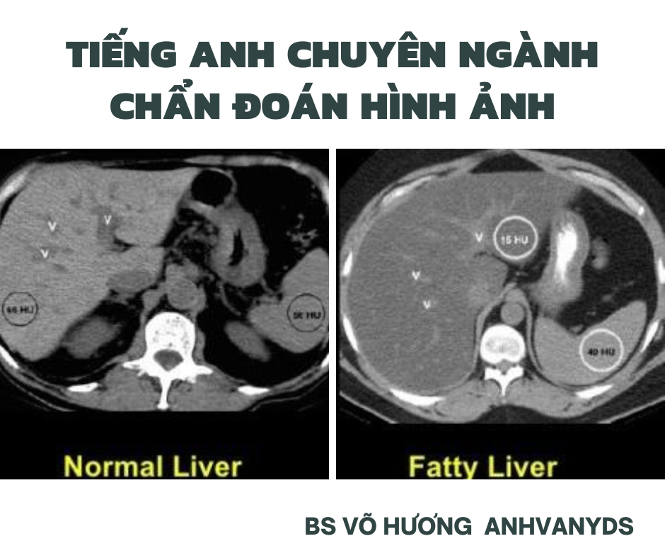
- Giảm tỷ trọng (Decreased attenuation) của gan ở hình ảnh trước tiêm thuốc (precontrast )và hình ảnh sau tiêm thuốc thì tĩnh mạch cửa portal venous phase imaging.
- Vùng gan bảo tồn không bị nhiễm mỡ (focal fatty sparing):
Không có tính chất giống với GNM lan tỏa.
Xuất hiện dưới dạng các vùng dạng bản đồ tăng tỷ trọng: hyperattenuating geographic regions)
PHẦN II: TẠM DỊCH
1. Plain radiograph (phim X quang không chuẩn bị)
Radiolucent liver sign: liver soft-tissue outline becomes difficult to appreciate.
Ultrasound (Siêu âm)
1.1.Steatosis manifests as increased echogenicity and beam attenuation. This results in:
- renal cortex appearing relatively hypoechoic compared to the liver parenchyma (normally liver and renal cortex are of a similar echogenicity)
- increased echogenicity relative to the spleen, when there is parenchymal renal disease
- absence of the normal echogenic walls of the portal veins and hepatic veins
- important not to assess vessels running perpendicular to the beam, as these produce direct reflection and can appear echogenic even in a fatty liver
- poor visualization of deep portions of the liver
- poor visualization of the diaphragm
1.2. Grading of diffuse hepatic steatosis on ultrasound has been used to communicate to the clinician about the extent of fatty changes in the liver.
2. GRADE (phân độ)
grade I: diffusely increased hepatic echogenicity but periportal and diaphragmatic echogenicity is still appreciable
grade II:diffusely increased hepatic echogenicity obscuring periportal echogenicity but diaphragmatic echogenicity is still appreciable
grade III: diffusely increased hepatic echogenicity obscuring periportal as well as diaphragmatic echogenicity
1.3. PRACTICAL POINTS (Các chú ý lâm sàng):
It is important not to fall into the pitfall that all diffusely echogenic livers are fatty, other pathologies may produce identical appearances, including cirrhosis.
Some suggest that visual grading of hepatic steatosis is subject to a wide interobserver and intraobserver variability.
3. Sonoelastography: can assess the degree of accompanying fibrosis by measuring tissue stiffness (FibroScan®, acoustic radiation force impulse).
There is also a histological three point scale for grading severity of non-alcoholic steatohepatitis. These two grading systems are not currently correlated.
4. CT (computed tomography): Cắt lớp vi tính.
- Diffuse steatosis reduces liver attenuation.
(Hepatic attenuation on CT, reflected by Hounsfield values, depends on a combination of factors including the presence or absence, as well as the phase, of IV contrast administration. Allowing for all these factors, the mean unenhanced attenuation value is around 55 HU. Several intrinsic liver pathologies can cause a diffuse change in liver attenuation with increased hepatic fat being the most prevalent.)
- On noncontrast CT, moderate to severe steatosis (at least 30% fat fraction) is predicted by:
relative hypoattenuation: liver attenuation lower than 10 HU less than that of spleen.
absolute low attenuation: liver attenuation lower than 40 HU.
- In comparison, contrast enhanced CT is poorly predictive of steatosis due to variation in both hepatic absolute enhancement and relative enhancement compared to spleen depending on contrast administration protocol, scan timing, and patient factors affecting contrast circulation . Nevertheless, some criteria for diffuse hepatic steatosis on contrast enhanced CT have been proposed:
liver-spleen differential attenuation (liver minus spleen) cutoffs ranging from less than -20 to less than -43 HU on portal venous phase, depending on injection protocol.
- Focal fatty sparing: (appearing as qualitatively hyperattenuating geographic regions) along the gallbladder fossa or periphery of segment
Nguồn tham khảo:
https://radiopaedia.org/articles/diffuse-hepatic-steatosis
BS VÕ HƯƠNG – ANHVANYDS.
Để lại một phản hồi Hủy