

Changes in oestrogen and progesterone
A pregnant woman’s progesterone levels are also very high. Among other effects, high levels of progesterone cause some internal structures to increase in size, including the uterus, enabling it to accommodate a full-term baby. It has other effects on the blood vessels and joints.
Changes in the uterus, cervix and vagina
After conception, the uterus provides a nutritive and protective environment in which the fetus will grow and develop. It increases from the size of a small pear in its non-pregnant state to accommodate a full-term baby at 40 weeks of gestation. The tissues from which the uterus is made continue to grow for the first 20 weeks, and it increases in weight from about 50 to 1,000 gm (grams). After this time, it doesn’t get any heavier, but it stretches to accommodate the growing baby, placenta and amniotic fluid. By the time the pregnancy has reached full term, the uterus will have increased to about five times its normal size:
The cervix remains 2.5 cm long throughout pregnancy. In late pregnancy, softening of the cervix occurs in response to increasing painless contractions of its muscular walls.
The vagina also becomes more elastic towards the end of pregnancy. These changes enable it to dilate during the second stage of labour, as the baby passes down the birth canal.
Pregnancy-related changes in posture and joints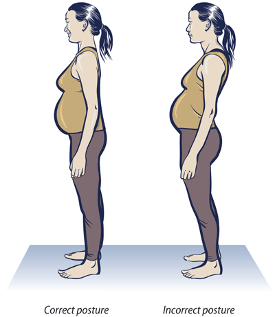

A pregnant woman’s entire posture changes as the baby gets bigger. Her abdomen transforms from flat or concave (dished) to very convex (bulging outwards), increasing the curvature of her back. The weight of the fetus, the enlarged uterus, the placenta and the amniotic fluid (the bag of waters surrounding the baby), together with the increasing curvature of her back, puts a large strain on the woman’s bones and muscles. As a result, many pregnant women get back pain. Too much standing in one place or leaning forward can cause back pain, and so can hard physical work. Most kinds of back pain are normal in pregnancy, but it can also be a warning sign of a kidney infection.
In addition, progesterone causes a loosening of ligaments and joints throughout the body. Pregnant women may be at greater risk of sprains and strains because the ligaments are looser, and because their posture has changed.
Changes in body weight during pregnancy
Continuing weight increase in pregnancy is considered to be one favourable indication of maternal adaptation and fetal growth. However, routine weighing of the mother during pregnancy is not now thought to be necessary, because it does not correlate well with pregnancy outcomes. For example, there can be a slight loss of weight during early pregnancy if the woman experiences much nausea and vomiting (often called ‘morning sickness’)
- About 2.0 kg in total in the first 20 weeks
- Then approximately 0.5 kg per week until full term at 40 weeks
- A total of 9 -12 kg during the pregnancy.
A lack of significant weight gain may not be a cause for concern in some women, but it could be an indication that the fetus is not growing properly. Doctors and midwives may refer to this as intrauterine growth restriction (IUGR) of the fetus.
Changes in the cardiovascular system

Blood volume
Blood volume (the total volume of blood in the circulation, measured in litres) increases gradually by 30-50 % in the pregnant woman, so by full term she has about 1.5 litres more blood than before the pregnancy. A higher circulating blood volume is required to provide extra blood flow through the placenta, so nutrients and oxygen can be delivered to the fetus. The increase in blood volume is caused by two changes:
- Increase in the volume of blood plasma (the fluid part of the blood).
- Increase in the number of red blood cells in the circulation.
The volume of blood plasma increases after about the sixth week of pregnancy. It reaches its maximum level of approximately 50% above non-pregnant values by the second trimester, and maintains this until full term.
The total volume of red cells in the circulation increases by about 18% during pregnancy, in response to the extra oxygen requirements made by the maternal, placental and fetal tissues. Red blood cells contain the oxygen-carrying substance called haemoglobin, which is rich in iron (see Box 7.1). Taking iron supplements during pregnancy can result in a much greater increase in red blood cells, up to 30% more than non-pregnant levels.
Blood pressure
Lower blood pressure is particularly common in early pregnancy. Many women report occasionally feeling dizzy in the first trimester, because less blood and less oxygen is being pumped to the brain. Progesterone can also cause a sudden larger relaxation in the blood vessels, resulting in an acute feeling of dizziness, or even a brief loss of consciousness (passing out).
Another cause of dizziness can result from lying flat on the back. This is more common after 24 weeks of pregnancy, but it can happen earlier during twin pregnancies, or conditions that increase the volume of amniotic fluid (waters surrounding the fetus). When a pregnant woman is lying flat on her back, the weight of her uterus and its contents compresses the large blood vessel (vena cava) leading from her lower body to the heart. When this blood vessel is squashed, the blood flow back to the heart is reduced, which in turn leads to a reduction in the blood flow out of the heart to the rest of the body.
Respiratory changes
During pregnancy, the amount of air moved in and out of the lungs increases by nearly 50% due to two factors:
- each breath contains a larger volume of air
- the rate of breathing (breaths per minute) increases slightly.
During pregnancy, many women find they get short of breath (cannot breathe as deeply as usual). This is because the growing baby crowds the mother’s lungs and she has less room to breathe. But if a woman is also weak and tired, or if she is short of breath all of the time, she should be checked for signs of sickness, heart problems, anaemia or poor diet. Get medical advice if you think she may have any of these problems.
Changes in the gastrointestinal system in pregnancy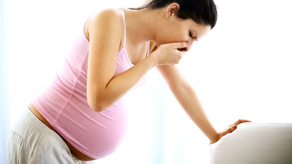

During pregnancy, the muscles in the walls of the gastrointestinal system relax slightly, and the rate at which food is squeezed out of the stomach and along the intestines is slowed down.
Many women also have nausea in the first months of pregnancy. A burning feeling, or pain in the stomach or between the breasts, is called indigestion (or ‘heartburn’, although the heart is not involved). It happens because as the pregnancy progresses, the growing baby crowds the mother’s stomach and pushes it higher than usual. The acids in the mother’s stomach that help digest food are pushed up into her chest, where they cause a burning feeling. This is not dangerous and usually goes away after the birth.
If the mother has difficulty with nausea or indigestion, advise her to eat small, frequent meals. The mother should not lie down flat for 1 to 2 hours after eating, because this may cause these symptoms.
Changes in the urinary system during pregnancy
Needing to urinate (pee) often is normal, especially in the first and last months of pregnancy. This happens because the growing uterus presses against the bladder. In late pregnancy, a woman often has to get up during the night to urinate, because fluid retained in the legs and feet during the day (oedema) is absorbed into the blood circulation when her legs are raised in bed. The kidneys extract the excess fluid and turn it into urine, so the bladder fills more quickly at night.
Changes in the breasts
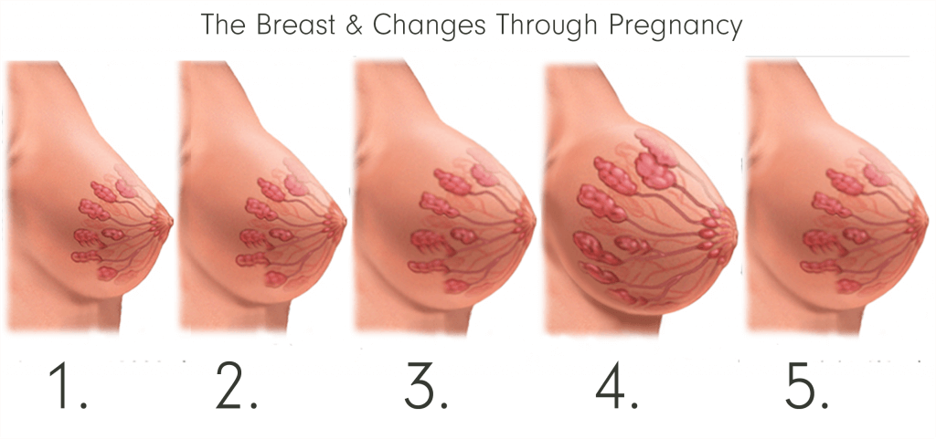

In early pregnancy, the breasts may feel full or tingle, and they increase in size as pregnancy progresses. The areola around the nipples (the circle of pigmented skin) darkens and the diameter increases. The Montgomery’s glands (the tiny bumps in the areola) enlarge and tend to protrude (stick out more). The surface blood vessels of the breast may become visible due to increased circulation, and this may give a bluish tint to the breasts.
By the 16th week (during the second trimester), the breasts begin to produce colostrum. This is the precursor of breastmilk. It is a yellowish secretion from the nipples, which thickens as pregnancy progresses. It is extremely high in protein and contains antibodies (special proteins produced by the mother’s immune system) that help to protect the newborn baby from infection. Near the end of pregnancy, the nipples may produce enough colostrum to make wet patches on the woman’s clothes. Reassure her that this is normal and a good sign. After the baby is born, colostrum is produced for about the first three days, before the proper milk begins to flow. Make sure that the mother breastfeeds the colostrum to her baby, so he or she gets all the nutrients and antibodies it contains.
Text source: http://www.open.edu
Tham khảo những bài giảng khác tại anhvanyds.com
Để lại một phản hồi Hủy