Reading
ANATOMY OF THE NIPPLE AND BREAST DUCT
Several important remarks on the nipple areolar complex NAC anatomy were made by Sir Astley Cooper [1840]. Over the last 160 years some of the anatomy knowledge acquired has changed since Cooper’s original work. For example, the glandular tissue is depicted as 15-20 lobes radiating out from the nipple, whereas Cooper stated that he observed up to 22 ducts leading to the nipple but considered that many of these ducts were not functional and that there were normally fewer than 12 patent ducts opening at the nipple.
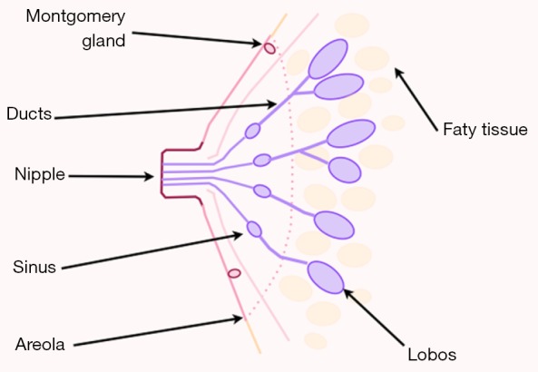
Actually, the breast is described as being composed of glandular and adipose tissue held together by a loose framework of fibers called Cooper’s ligaments. Histological studies demonstrate that the lobes are composed of lobules, which consist of clusters of alveoli containing mammary secretory epithelial cells. The alveoli are connected to very small ducts that join to form larger ducts draining the lobules. These larger ducts finally merge to a unique duct for each lobe. Under the areola, this single duct is depicted as widening into a lactiferous sinus before narrowing at the base of the nipple and terminating at its orifice on the surface of the nipple. Adipose tissue of the breast is situated between lobes rather than within lobules.
Nipple abnormality
These anomalies could be classified into multiple conditions.
-
Accessory nipple. One to five percent of the population, equal incidence in male and females. Usually found in the inframammary region, and are prone to the same diseases as normal nipples. Excision is only indicated for cosmetic purposes, discomfort during menstruation, anxiety, pain, or restriction of arm movement .
-
Athelia is characterized by the congenital absence of the nipple areolar complex with the presence of breast tissue . This condition can be inherited by autosomal dominant inheritance, or as a part of a syndrome (e.g., Poland’s).
-
Amastia is the complete absence of breast structures.
-
Amastia is the absence of breast tissue with preservation of the NAC.
-
Inverted nipple is a condition where the nipple, instead of pointing outward, is retracted into the breast. In some cases, the nipple will be temporarily protruded if stimulated, but in others, the inversion remains regardless of stimulus. Women and men can have inverted nipples. Most common nipple variations that women are born with are caused by short ducts or a wide areola muscle sphincter .
Anatomy of NAC
The NAC comprises two basic structures: the areola and the nipple
The areola has a round shape and a varying size, on average 3 to 6 centimeters, normally situated around the forth rib level. It has sebaceous glands that make projections on its surface, forming tubercle of morgani, or areolar glands, which during pregnancy become enlarged giving rise to tubercles of Montgomery.
Some authors believe that such structures represent accessory mammary glands and it has been observed milk secretion output with manual expression of these tubers (this supposition leads some surgeons to prefer the use of the skin sparing mastectomy instead of nipple or areola sparing mastectomies because of oncological concerns).
In the center of the areola emerges a papillar cylindrical formation varying in size, averaging 10 to 12 millimeters (mm) wide by 9 to 10 mm in height. Its skin is similar to the areola, but has no sebaceous glands. It has 10 to 20 corresponding pores as the output of the milk ducts.
The NAC has no subcutaneous tissue. The skin of the nipple rests on a thin layer of smooth muscle, areolar muscle fibers which are distributed in two directions: radial and circular. The muscle of Sappey responsible for circular fibers and the muscle of Meyerholz, formed by the radial fibers.
The areolar muscle is continued in the papilla with longitudinal and circular fibers surrounding the milk ducts along with connective tissue support. Its contraction is responsible for the ejection of secretion in the milk sinuses and for the telotism of the papilla, “mimicking an erection”.
Below the areolar muscle there is a thin layer of fat which disappears as it approaches the papilla. In this pre-mammary fat tissue layer vessels are found running on radiated sense.
Source: Anatomy of the nipple and breast ducts
FURTHER READING
NIPPLE VARIATIONs
https://www.breastfeeding-problems.com/nipple-variations.html
Xem thêm những bài giảng khác tại: https://anhvanyds.com/
Để lại một phản hồi Hủy